
Fibrous membranes of poly(3-hydroxybutyrate-co-3-hydroxyhexanoate) with ceftriaxone: An investigation focused on wound healing applications
Antônio Schandler Ferri , Maria Eduarda Araújo Ribeiro, Marina Rangel Ferro Pereira, Fabrício Moreira Almeida, Theresa de Moura Monteiro, Fernanda Antunes, Jose Gregório Cabrera Gomez, Ruben J. Sanchez Rodriguez
Vol. 20., No.10., Pages 1089-1103, 2026
DOI: 10.3144/expresspolymlett.2026.79
DOI: 10.3144/expresspolymlett.2026.79
Corresponding author: Maria Eduarda Araújo Ribeiro 
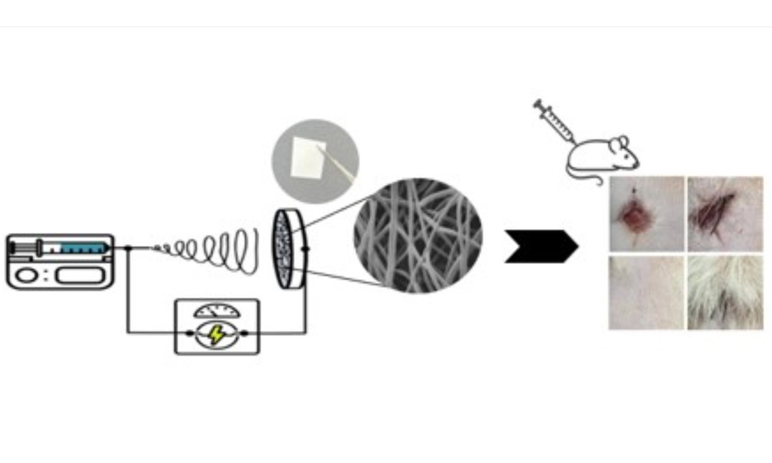
GRAPHICAL ABSTRACT
ABSTRACT
Conventional wound dressings, such as gauze, primarily act as physical barriers and provide limited support for tissue regeneration. In this study, electrospun poly(3-hydroxybutyrate-co-3-hydroxyhexanoate) (P(3HB-co-3HHx)) membranes were developed as multifunctional wound dressings combining a biomimetic fibrous structure with localized antibiotic delivery. Pristine and ceftriaxone (CFX)-loaded membranes were fabricated by electrospinning (15 kV, 1 mL/h, 20 cm, 6 h), yielding membranes approximately 300 μm thick. SEM revealed an interconnected fibrous architecture resembling the skin extracellular matrix, with mean fiber diameters of 1.38 ± 0.48 and 1.45 ± 0.57 μm for pristine and CFX-loaded membranes, respectively. Both membranes exhibited suitable wettability, water vapor transmission, and water sorption, while CFX incorporation reduced the water contact angle by approximately 17°, producing a less hydrophobic surface. CFX localization on the fiber surface resulted in an initial burst release, followed by diffusion-controlled release over 120 h. The membranes showed favorable in vitro and in vivo biological performance, with no cytotoxic effects and enhanced wound closure compared with commercial dressings. Overall, P(3HB-co-3HHx)/CFX membranes show potential as bioactive wound dressings combining extracellular-matrix-like architecture and localized antibiotic delivery.
RELATED ARTICLES
Delia Mihaela Raţă, Anca Niculina Cadinoiu, Luiza Mădălina Grădinaru, Paul Codrin Fuioaga, Gabriela Vochita, Christelle Delaite, Leonard Ionuţ Atanase
Vol. 19., No.3., Pages 233-245, 2025
DOI: 10.3144/expresspolymlett.2025.18
Vol. 19., No.3., Pages 233-245, 2025
DOI: 10.3144/expresspolymlett.2025.18

Curcumin (CRC) is a natural active principle with important anti-inflammatory, antioxidant, antibacterial, and antitumor properties but has some limitations, such as poor bioavailability, low water solubility, and rapid metabolism. To preserve CRC’s benefits and eliminate its limitations, novel CRC-loaded core-shell electrospun nanofibers were designed. The nanofibers were prepared by co-axial electrospinning method using poly(vinyl alcohol)/CRC as core and poly(vinyl alcohol)/sodium alginate as shell. Polymer coating protects the CRC, increasing its stability. The swelling degree of CRCloaded nanofibers at pH 5.4 was around 326% higher than at pH 7.4 (297%) due to the repulsions of the anion-anion COO–groups. The release efficiency of CRC at pH 7.4 was 81%, while at pH 5.4 was about 96%. In the first 6 h, there was a slower release of CRC from the nanofibers in both acidic and slightly alkaline environments. Nanofibers showed good hemocompatibility, the values being between 2.36–3.22% after the first 90 min of contact, and after 180 min of treatment, the degree of erythrocyte lysis was between 3.78 and 4.93%. Cell viability assay on V79-4 Chinese hamster fibroblasts demonstrated that treatment with free CRC led to a value of 39% whereas for CRC-loaded nanofibers, the cell viability value increased to 59.66%. The results of the present study indicated that CRC-loaded electrospun nanofibers can have great potential for biomedical applications.
Aleksandra Sierakowska-Byczek, Julia Radwan-Pragłowska, Łukasz Janus, Tomasz Galek, Karol Łysiak, Mirosław Tupaj, Dariusz Bogdał
Vol. 18., No.8., Pages 819-834, 2024
DOI: 10.3144/expresspolymlett.2024.61
Vol. 18., No.8., Pages 819-834, 2024
DOI: 10.3144/expresspolymlett.2024.61
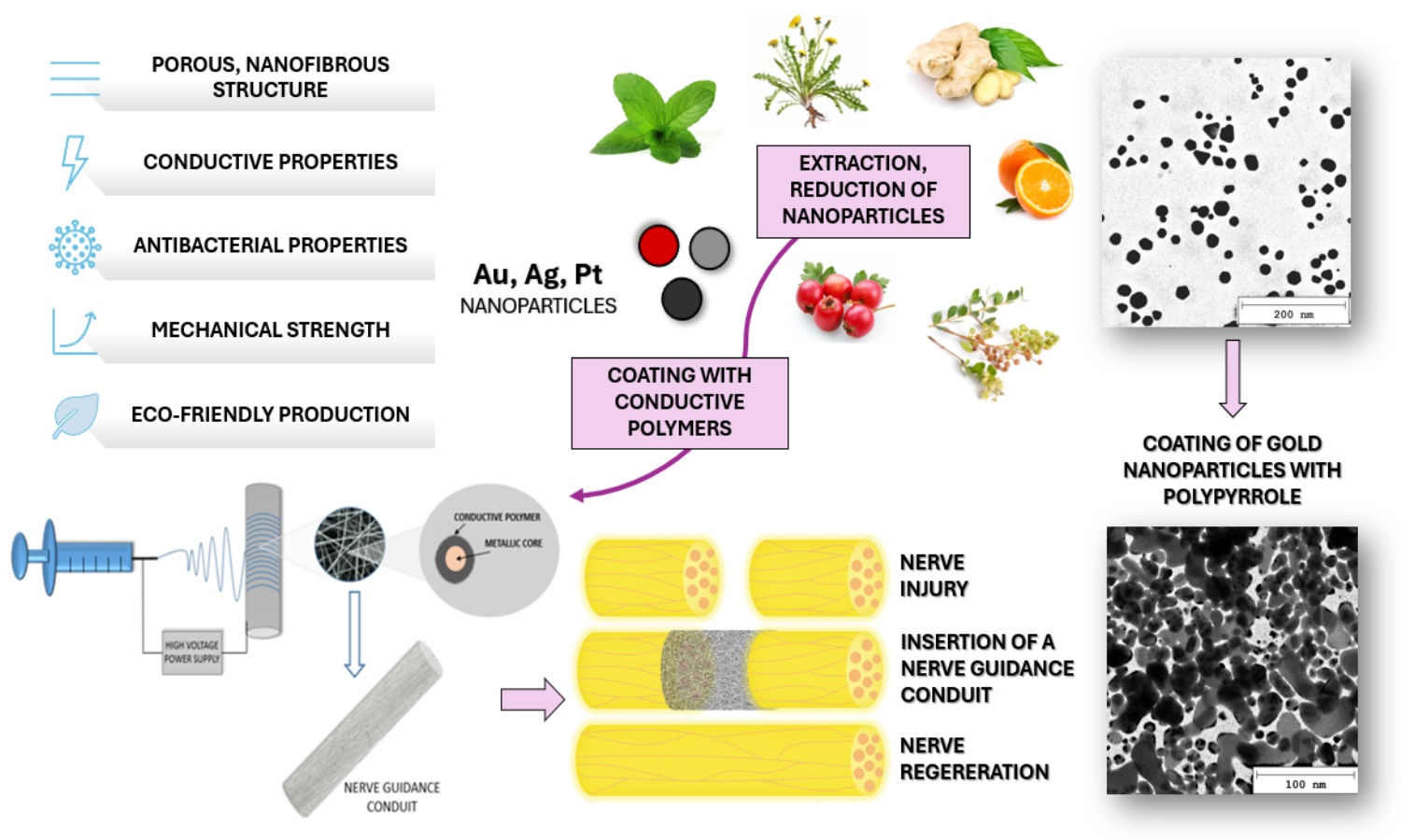
Appropriate protection and guiding are crucial during peripheral nerves repair. New generation nerve guidance conduits (NGCs) should not only provide mechanical support for the damaged nerve but also support healing processes. One of the most promising tissue regeneration applications is fibrous biomaterials since they are characterized by high porosity, flexibility, and strength. Additionally, they enable cell adhesion and proliferation. In this study, novel fibrous nanocomposites were obtained by applying the electrospinning technique, using polylactic acid (PLA) as a polymeric matrix which was further modified with metallic nanoparticles coated with conductive polymers. Such an approach resulted in the obtainment of biomaterials with a potential ability to conduct nerve impulses. The chemical structure of the obtained composites, as well as the morphology of ready products and separate nanocomponents, were investigated using Fourier-transform infrared spectroscopy (FTIR), transmission electron microscope (TEM) and scanning electron microscope (SEM) techniques. Furthermore, conductive and swelling properties in various media were determined. Finally, biomaterials were confirmed to be non-cytotoxic to L929 mouse fibroblasts and 1321N1 human glial cells. Based on the presented results, it can be concluded that nanofibrous nerve guidance conduits have all the key properties in the process of peripheral nerve regeneration and may constitute an important step in novel NGCs development.